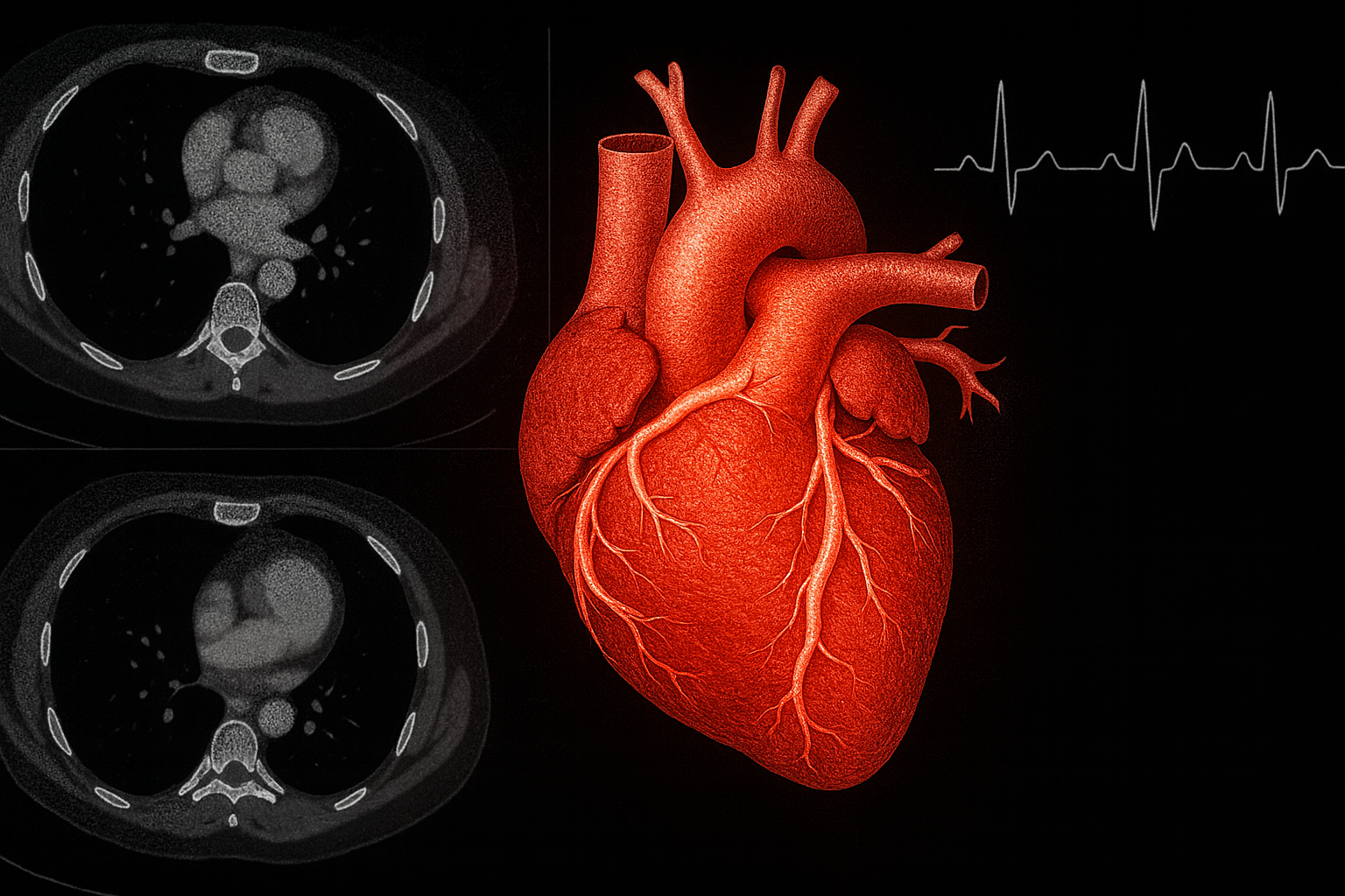
Advanced cardiovascular imaging plays a pivotal role in diagnosing, risk-stratifying, and managing a spectrum of cardiac conditions, from ischemic heart disease to cardiomyopathies and valvular disorders. However, rapid technological evolution—spanning echocardiography (echo), cardiovascular computed tomography (CCT), cardiovascular magnetic resonance (CMR), and nuclear cardiology—demands rigorous, standardized training to ensure competency and patient safety. The 2025 Advanced Training Statement on Advanced Cardiovascular Imaging, jointly authored by the American College of Cardiology (ACC), American Heart Association (AHA), American Society of Echocardiography (ASE), American Society of Nuclear Cardiology (ASNC), Society of Cardiovascular Computed Tomography (SCCT), and Society for Cardiovascular Magnetic Resonance (SCMR), and issued by the ACC Competency Management Committee, addresses this need by updating prior frameworks to reflect contemporary practice.
This comprehensive report delineates core competencies at Level II (independent performance) and Level III (advanced expertise, including teaching and research) for each modality. For echo, trainees must achieve proficiency in 3D imaging, strain analysis, and interventional guidance, with a minimum of 150 supervised studies. CCT training emphasizes coronary plaque characterization and structural interventions, requiring 150 cases including 50 CT angiograms. CMR competencies cover perfusion, viability, and congenital assessments, mandating 100 studies with contrast-enhanced protocols. Nuclear cardiology focuses on PET/SPECT hybrid imaging and theranostics, with 100 interpretations, including 50 attenuation-corrected scans. Cross-cutting skills include radiation safety, contrast management, multimodality fusion, and ethical considerations like cost-effectiveness.
Training pathways recommend 12-24 months of dedicated fellowship for Level II, extendable to 36 months for Level III, incorporating simulation labs, AI-driven analytics, and interprofessional collaboration. New emphases include health equity (e.g., addressing disparities in imaging access for underserved populations), quality metrics via registries like IMAGE and IC3, and continuous professional development through maintenance of certification modules. Assessment tools encompass direct observation, logbooks, and milestone evaluations aligned with the ACGME/ABIM frameworks.
By harmonizing expectations across societies, this statement fosters a workforce adept at value-based imaging, reducing overuse while enhancing outcomes. As lead author Dr. Pamela Douglas notes, “These competencies empower imagers to lead in an era of precision medicine.” Future iterations may incorporate emerging technologies like photon-counting CT and machine learning algorithms. This document not only guides program directors and trainees but also informs accreditation bodies, ultimately elevating cardiovascular care standards globally.
Link: https://www.ahajournals.org/doi/10.1161/HCI.0000000000000088